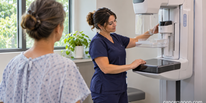
A mammogram after lumpectomy looks for changes in the remaining breast tissue. After breast-conserving surgery and radiation, the first examination is often scheduled about 6 to 12 months after radiation ends. Confirm your timing with the team that manages your follow-up.
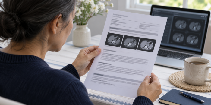
CT Scan After Colon Cancer Treatment: How to Read the Report
CancerSnoop Follow-Up Guide
CT Scan After Colon Cancer Treatment: How to Read the Report
A CT scan after colon cancer treatment looks for changes within the body. Learn how the findings, comparison scans and impression fit into your follow-up plan.
| Test Name | Follow-up CT report |
|---|---|
| Test Type | Imaging |
| What It Measures | Cross-sectional images of the body regions included in the examination. |
| Used For | Evaluate changes during follow-up and investigate specific findings or symptoms. |
Begin with the reason for the examination
A CT scan after colon cancer treatment should be read in the context of why it was ordered. A planned surveillance examination asks a different question from a scan arranged because of a new symptom or an earlier uncertain finding. The clinical history section may summarize that reason. If important treatment is missing from the history, mention it to your team. The person interpreting the examination benefits from knowing the relevant operation, treatment and question that prompted the scan. [1]
Check which regions and technique were used
The technique section explains the body regions examined and whether contrast was given. A report for the abdomen and pelvis does not describe the chest unless the chest was also included. Contrast can make structures more distinct, and the timing and type of examination affect what can be assessed. Before a future appointment, tell the imaging service about a previous contrast reaction or kidney problem. Ask the ordering team if you are unsure why the scan was arranged with or without contrast. [2]
Find the comparison section before interpreting a measurement
The comparison section identifies previous imaging available to the radiologist. An older scan can show whether a small area was already present, while a scan performed after treatment may establish a new baseline. If your imaging has been performed at different hospitals, ask whether the actual earlier images were available. A written report alone may provide less detail for comparison. When you see a measurement, read it together with the description of change and the radiologist's interpretation, rather than judging the number in isolation. [1]
Read findings and impression together
The findings describe observations by organ or region. The impression brings together the main conclusions and recommendations. A finding can be described in detail without becoming the most important issue in the impression. Conversely, a short recommendation for another examination may be the key next step. Look for whether the radiologist has identified a likely explanation, expressed uncertainty or recommended comparison or follow-up. If the impression is unclear to you, ask the clinician what question remains unanswered and how they plan to answer it. [1]
What do stable and indeterminate mean?
Stable means the finding has not changed in the way described over the comparison interval. Indeterminate means the examination has not established what the finding is. These words describe different aspects of the assessment. An indeterminate area can also be stable, and its significance depends on features, history and the available comparison. Ask what degree of confidence the radiologist expressed and whether the recommendation is routine follow-up or a more focused examination. The report should lead to a plan you can understand. [1]
Why might MRI follow a CT?
A CT can identify a liver finding that needs further characterization. In that setting, MRI may provide different views and tissue information. Ordering it means the team is seeking more information about a particular finding. It does not by itself establish that the finding is cancer. Ask what the MRI is expected to clarify, when it should happen and who will explain both results together. The separate liver-lesion article in this series describes that process without assuming every liver spot has the same cause. [4]
What should you do after reading the report online?
Write down the exact phrase you want explained and the date of the scan. Ask which part of the report changes the follow-up plan, which earlier images were compared and whether any recommended examination has been ordered. A portal may release the report before your clinician has discussed it with you, so arrange that conversation. Keep the agreed next step with your appointment record. If you have symptoms needing urgent attention, use the instructions from your care team rather than waiting for a routine results appointment. [1]
What does a reassuring examination actually tell you?
It tells you what the team could assess at that time using that examination. Keep the scheduled follow-up and discuss new concerns with the appropriate clinician. If the report recommends more information, ask whether the uncertainty affects the conclusion or refers to a separate finding. The aim of reading the report is to understand the next decision. You do not need to translate every technical term to ask whether the findings change your care or whether there is a task you need to complete before the next visit. [3]
Sources and References
- How to Read Your Abdominal and Pelvic CT Report
- Computed Tomography (CT) Scans and Cancer
- Living as a Colorectal Cancer Survivor
- Body MRI
Evidence coverage: guidance and educational sources were checked for this guide. Recommendations are described within their clinical scope. This educational article does not replace your care team’s interpretation of your results.
Continue Exploring