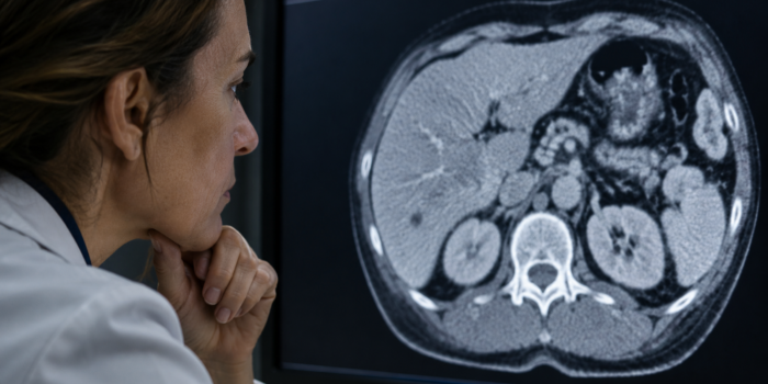
A mammogram after lumpectomy looks for changes in the remaining breast tissue. After breast-conserving surgery and radiation, the first examination is often scheduled about 6 to 12 months after radiation ends. Confirm your timing with the team that manages your follow-up.
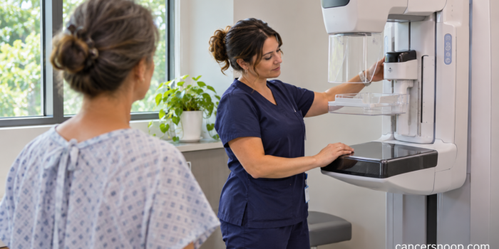
Mammogram Callback: Why You Were Asked to Return
CancerSnoop Test Guide
Mammogram Callback: Why You Were Asked to Return
A phone call after screening can leave you wondering what the team saw and how quickly you need to return.
| Test Name | Diagnostic mammogram after screening |
|---|---|
| Test Type | Imaging |
| What It Measures | The shape, margins and distribution of an area that was unclear on screening, or tissue that was not fully captured. |
| Used For | Resolve a specific screening question before deciding whether routine screening, monitoring or sampling is appropriate. |
| Typical / Reference Range | The assessment category and the specific area to recheck. |
What the original pictures could not settle
Screening images are designed to give a broad view of the breasts. When a region looks different from the surrounding tissue, the radiologist may need images taken from a different angle or with pressure focused over that spot. Sometimes overlapping tissue separates on those images and the concern disappears. Other times the finding remains visible and the radiologist needs ultrasound or another study. A return appointment may also be requested when prior images are unavailable or positioning needs to be repeated. [1] [2]
What happens at the mammogram callback
A diagnostic visit starts with a specific question. Ask the team which breast and which area prompted the call. The technologist may take targeted images and show them to a radiologist while you are still there. An ultrasound can be added when it is useful for examining an area or distinguishing a fluid-filled cyst from a solid mass. Ultrasound may contribute little when the concern is a pattern of tiny calcium deposits seen only on mammography. The team chooses the views that best answer the question. [2] [3]
How to read the result you get next
Check both the assessment and the recommendation. If the area is resolved as normal or benign, the report can recommend routine screening. A finding judged probably benign may require imaging at a shorter interval. A suspicious finding can lead to a biopsy recommendation. These are decisions about what the images support and what needs to happen next. If a separate appointment is required, ask which office will schedule it and when you should expect to hear back. [1]
Why old images can help
A prior report may say that the previous screening examination was normal without showing the exact shape and location of the tissue the radiologist wants to compare. The images themselves give the team more information. Tell the center where and when your previous studies were done. Ask whether it can request the files or whether it needs you to arrange the transfer. If you have noticed a lump, nipple change or other symptom, describe it when scheduling so the diagnostic team knows about it.
What to write down after the visit
Record the final assessment, the next test if one is needed and the date or interval the report recommends. A practical question is, “Did today's imaging resolve the finding from screening?” If the answer is no, ask what remains uncertain and how it will be resolved. If a different finding was discovered, ask the team to point out which recommendation belongs to each area. This helps when the report lists more than one finding.
What if I have already had an ultrasound?
Tell the imaging center when it was done and ask whether it examined the same site. A prior ultrasound that looked at a different area may leave the mammography question open. The center can determine whether to use those images, obtain additional mammographic views or perform a new targeted ultrasound.
Sources and References
Continue Exploring