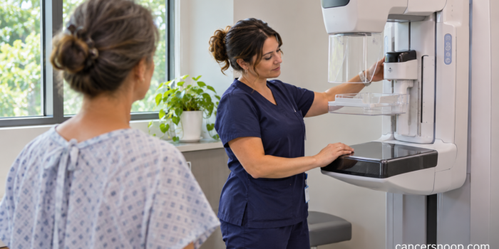
A mammogram after lumpectomy looks for changes in the remaining breast tissue. After breast-conserving surgery and radiation, the first examination is often scheduled about 6 to 12 months after radiation ends. Confirm your timing with the team that manages your follow-up.
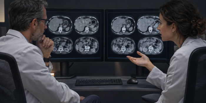
A New Liver Spot After Colon Cancer: What Happens Next?
CancerSnoop Follow-Up Guide
A New Liver Spot After Colon Cancer: What Happens Next?
A liver lesion after colon cancer needs assessment in context. The scan appearance, older images and sometimes a liver MRI help the team decide what the finding means.
| Test Name | Liver lesion characterization |
|---|---|
| Test Type | Imaging |
| What It Measures | The appearance of a focal liver finding using relevant imaging and comparison studies. |
| Used For | Clarify an uncertain liver finding after colon cancer and guide further evaluation. |
What does the word lesion tell you?
A liver lesion after colon cancer is an area that looks different from surrounding liver tissue on an examination. The word itself does not identify the cause. A report may offer a likely explanation, describe the area as indeterminate or recommend another examination. Start with the actual wording rather than replacing it with a diagnosis. If someone calls it a spot in conversation, ask what term the radiologist used and whether a specific explanation was given. [1]
Does new mean it has definitely appeared since the last scan?
The comparison matters. The radiologist may be describing a finding absent from an earlier examination, or one that is better seen using a different technique. Ask which earlier images were compared and whether the finding was visible in retrospect. If the prior scan came from another hospital, the team may need those images. A comparison can change the interpretation or recommendation. The date, body region and technique of the earlier examination provide context that the single word new cannot convey. [1]
What features can the team assess?
The radiologist considers the size, shape, internal appearance and how the area behaves with contrast when that information is available. Some appearances support a benign explanation, while others require closer assessment. Your history of colon cancer also matters because the liver is a relevant site to assess during follow-up. Neither history nor size alone supplies the answer. Ask whether the report names a likely cause, lists possibilities or simply says that the examination could not characterize the lesion. [3]
Why might the next step be a liver MRI?
MRI can provide additional tissue information and a more focused assessment of a liver finding. RadiologyInfo's CT report guide gives an example of an indeterminate liver lesion in someone with colorectal cancer for which MRI is recommended. That is an example of how an unresolved question can lead to another test. It is not a rule for every lesion. Ask what protocol the team plans to use and whether the result could alter your treatment or follow-up. [2]
Would a biopsy always give the answer sooner?
Biopsy takes tissue and can be useful in selected situations, but the choice depends on the clinical question and the imaging already available. Some lesions can be characterized through imaging. In other cases, a multidisciplinary review may determine whether tissue is needed and what result would change care. Ask why the team recommends imaging, interval follow-up or biopsy in your situation. The reason is more informative than assuming one of those options is always the quickest or best route. [3]
How should you handle the time between examinations?
Before leaving the appointment, confirm whether an additional test was ordered, the intended timing and how you will receive the result. If the referral is not visible or the booking has not arrived, contact the named service. Keep the wording of the finding and the scan date in one place so that you can explain why you are calling. You can also ask whether the case needs review by specialists who routinely assess liver findings in people with colorectal cancer. [1]
What if further assessment suggests a metastasis?
Then the conversation changes from characterizing the finding to deciding what treatment options fit the whole situation. The team considers the number and location of liver findings, other disease sites, previous treatment and your health. This article cannot assign those options from a radiology phrase alone. Ask whether all relevant images and treatment records have been reviewed together. A clear result discussion should state what is known, what remains uncertain and what decision the next investigation is intended to support. [5]
What if the report uses a liver-specific score?
Some liver reporting systems address particular diseases and risk groups. A score developed to assess primary liver cancer in an at-risk population should be interpreted in its own setting. Ask which system was used and whether it applies to the finding being evaluated after colon cancer. If you read general liver information online, bring the question back to your team before applying its categories to yourself. A colorectal metastasis assessment and an assessment for primary liver cancer can involve different questions and different reporting language. [4]
Sources and References
- How to Read Your Abdominal and Pelvic CT Report
- Body MRI
- Management of Incidental Liver Lesions on CT: A White Paper of the ACR Incidental Findings Committee
- How to Read Your Liver Imaging Report using LI-RADS
- Colon and Rectal Cancer Follow-Up Care Expanded Version
Evidence coverage: guidance and educational sources were checked for this guide. Recommendations are described within their clinical scope. This educational article does not replace your care team’s interpretation of your results.
Continue Exploring