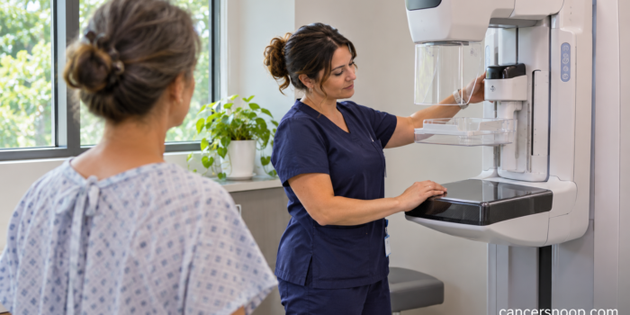
A mammogram after lumpectomy looks for changes in the remaining breast tissue. After breast-conserving surgery and radiation, the first examination is often scheduled about 6 to 12 months after radiation ends. Confirm your timing with the team that manages your follow-up.
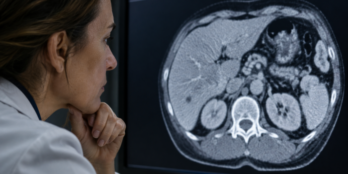
Too Small to Characterize on a Cancer Scan: What Does It Mean?
CancerSnoop Follow-Up Guide
Too Small to Characterize on a Cancer Scan: What Does It Mean?
Too small to characterize means an examination could not reliably identify a tiny finding. In cancer follow-up, the next step depends on its location, appearance, prior scans and clinical history.
| Test Name | Assessment of a finding described as too small to characterize |
|---|---|
| Test Type | Imaging |
| What It Measures | A small imaging finding whose nature cannot be reliably established on the examination. |
| Used For | Explain a radiology limitation and decide whether comparison or further imaging is appropriate. |
What information is missing when the report uses this phrase?
Too small to characterize means the examination shows a small area but cannot reliably determine its nature. The radiologist is distinguishing detection from characterization. Seeing a tiny spot and knowing what it is are separate achievements. The phrase may appear in a report about an organ such as the liver, and it needs to be read with the surrounding sentences. Ask whether there were any descriptive features, a likely explanation or a recommendation attached to it. [1]
Why can an examination detect something without identifying it?
An image has practical limits. A very small finding may not provide enough detail to assess its internal structure or the way it behaves with contrast. The technique used for a broader surveillance question may also differ from a focused examination of one organ. This is why another imaging method or comparison can be useful in selected cases. Ask what the limitation was in your examination. The answer helps explain why a follow-up recommendation was made. [1]
How can older scans help?
An earlier image may show the same area and allow the radiologist to describe whether it has changed. The length of the comparison interval and similarity of the examinations matter. Ask whether the prior images were available and which dates were compared. If the earlier examination was performed elsewhere, find out how the imaging service can obtain it. A written summary of an old scan and the actual images are different resources, and your team can advise what it needs for review. [1]
Why does cancer history change the discussion?
A person's history affects how a small finding is assessed. Guidance for an incidental liver finding in someone without known cancer may not fit a person undergoing colon cancer surveillance. The organ, the previous cancer and the purpose of the examination need to be considered together. Avoid applying a size threshold from a general information page to your report. Ask whether the finding is covered by a specific recommendation for your clinical setting and which features determine that recommendation. [2]
When might another scan be recommended?
The team may seek better characterization using a different method, or monitor the area over an agreed interval. For a liver finding, MRI is one possible way to obtain more information. The American College of Radiology's incidental liver guidance uses risk and imaging features to guide management, but its algorithm has a defined scope. It should not be converted into a universal timetable for every cancer patient. Your clinician should explain why the proposed examination and timing fit your case. [3]
What can you ask without having to interpret the images yourself?
Ask where the finding is, whether it was seen before and whether it looks suspicious or is simply unresolved. Then ask what result from another examination would change management. If follow-up is recommended, confirm who orders it and who will review the result. Those questions turn a vague phrase into a concrete plan. Keep the agreed plan in your follow-up record, particularly if your oncology and imaging appointments are at different facilities. [1]
How does the phrase fit with the rest of the report?
Read the impression and recommendation together with the detailed finding. A report may describe several observations with different levels of significance. The phrase alone cannot establish that recurrence is present or that the area is harmless. Ask your team which observations affect the plan now and which will be reassessed later. If the wording changes on a future report, compare the explanations with your clinician rather than assuming a changed adjective necessarily represents a biological change. [1]
Is there one size below which a finding can be ignored?
There is no universal size rule that answers every cancer follow-up question. The organ, appearance and history matter together. A recommendation designed for a small incidental liver finding in a low-risk adult cannot simply be transferred to a patient with a previous cancer that can spread to the liver. Ask your team to explain the applicable context. If they recommend watching an area, ask what would trigger a different examination and who is responsible for ensuring the planned reassessment takes place. [2]
Sources and References
- How to Read Your Abdominal and Pelvic CT Report
- Management of Incidental Liver Lesions on CT: A White Paper of the ACR Incidental Findings Committee
- Body MRI
Evidence coverage: guidance and educational sources were checked for this guide. Recommendations are described within their clinical scope. This educational article does not replace your care team’s interpretation of your results.
Continue Exploring