
More testing is not necessarily better surveillance. Evidence suggests that very frequent CEA testing and imaging after colorectal cancer treatment does not automatically improve outcomes. CEA works best as one part of the wider follow-up picture.
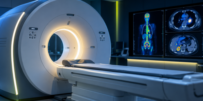
PET Scan in Cancer: What It Shows and What the Results Mean
CANCERSNOOP TEST GUIDE
A PET scan in cancer care can show where tissue is unusually active. It cannot tell you by itself whether that activity is cancer.
PET/CT can be extremely useful for staging cancer, investigating possible recurrence and assessing treatment response. But areas that “light up” are not automatically cancer, and cancer does not always light up.
The short answer
A PET scan is an imaging test that can show metabolic activity inside the body.
In cancer imaging, PET is commonly combined with CT. This is called PET/CT.
The PET part shows areas of increased tracer uptake, while the CT part helps doctors see exactly where those areas are located anatomically.
PET/CT can help answer important questions—but it is not a simple cancer detector.
✓
PET can help show
Where metabolically active disease may be present.
Depending on the cancer and clinical situation, PET/CT can contribute to staging, restaging, recurrence assessment and evaluation of treatment response.
!
PET cannot prove
That every bright area is cancer.
Infection, inflammation and normal activity in some tissues can also produce increased tracer uptake.
What is a PET scan in cancer care?
PET stands for positron emission tomography.
Unlike imaging that mainly shows anatomy, PET can provide information about biological activity in tissues.
For many cancer PET scans, a small amount of radioactive tracer called 18F-fluorodeoxyglucose (FDG) is used.
FDG behaves similarly to glucose. Tissues that take up more glucose may accumulate more FDG and become more visible on the PET images.
In plain language
PET is looking for patterns of metabolic activity—not simply taking a photograph of cancer.
Why does cancer sometimes “light up” on PET?
Many cancer cells use glucose at a higher rate than surrounding normal tissues.
When FDG accumulates in these cells, the area may show increased uptake on the PET scan.
This is why people often hear phrases such as:
• “the lesion is FDG-avid”
• “increased metabolic activity”
• “increased tracer uptake”
• “hypermetabolic lesion”
These phrases describe what the scan detected. They are not automatically a diagnosis of cancer.
What is PET/CT?
Modern cancer imaging often combines PET and CT in the same examination.
PET
What is active?
PET provides information about the distribution and intensity of tracer uptake.
CT
Where is it?
CT provides anatomical detail that helps locate and characterize what is seen on PET.
Combining the two can help doctors connect metabolic activity with a specific lymph node, organ, mass or other anatomical structure.
What is a PET scan used for in cancer?
The role of PET/CT varies by cancer type and clinical situation. It is not automatically required for every person with cancer.
Staging
PET/CT can help determine how far some cancers have spread and whether metabolically active disease is present in additional locations.
Restaging and suspected recurrence
PET/CT may help investigate whether disease has returned or progressed when recurrence is suspected.
Treatment response
Changes in metabolic activity can sometimes help doctors assess how disease is responding to treatment.
Does a bright spot on PET mean cancer?
No.
This is one of the most important limitations of FDG PET imaging.
FDG uptake is not specific to cancer.
Inflammation and infection can also increase glucose metabolism and produce areas of increased uptake.
Normal organs and tissues can also show expected physiological FDG activity.
IMPORTANT
“FDG-avid” does not mean “confirmed cancer.”
It means the area has taken up more FDG. Doctors still need to interpret why that happened using the location, CT appearance, clinical history, previous imaging and sometimes additional testing or biopsy.
Can cancer be present without showing on PET?
Yes.
A PET scan that does not show abnormal FDG uptake does not guarantee that cancer is absent.
There are several reasons disease may be difficult to detect.
Very small lesions can fall below the effective resolution of the scan. Some tumors also have relatively low FDG uptake, making them less conspicuous on FDG PET.
Timing, blood glucose, recent treatment and technical factors can also influence image quality and interpretation.
DON’T MISS THIS
A negative PET scan does not equal “no cancer.”
PET is powerful, but it has detection limits. The meaning of a negative scan depends on the cancer type, lesion size, treatment history and the other available evidence.
What is SUV on a PET scan?
You may see the abbreviation SUV in a PET report.
SUV stands for standardized uptake value. It is a quantitative measurement used to describe tracer uptake in tissue.
Higher SUV values can occur in cancer, but there is no universal SUV number that separates cancer from non-cancer in every patient, organ and tumor type.
SUV can also be influenced by technical and biological factors, which is why the number must be interpreted as part of the complete examination.
Key point
There is no single SUV cutoff that can independently tell you: “below this is benign, above this is cancer.”
What does “metabolic response” mean?
PET/CT can sometimes be used to evaluate how metabolic activity changes during or after treatment.
If previously active lesions show substantially less FDG uptake, the report may describe a metabolic response.
If abnormal uptake is no longer identified under the relevant response criteria, terms such as complete metabolic response may be used.
That wording needs to be interpreted carefully.
It describes what can be detected metabolically on the scan. It does not necessarily prove that every cancer cell in the body has disappeared.
PET scan vs CT scan: what is the difference?
They provide different kinds of information.
CT
Anatomy
Shows structures, lesion size, shape, location and anatomical changes.
PET
Metabolic activity
Shows the distribution of a radioactive tracer and areas with altered metabolic activity.
PET/CT
Both together
Combines metabolic information with anatomical localization in a single examination.
Why might PET and CT seem to disagree?
Because they are measuring different things.
A lesion may remain visible anatomically on CT even after its metabolic activity has fallen substantially.
In another situation, PET may show abnormal metabolic activity in an area where the structural change on CT is subtle.
This does not automatically mean one test is right and the other is wrong. Doctors interpret the two types of information together.
Can PET detect very small metastases?
Not reliably in every situation.
PET has spatial-resolution limits. Very small deposits may contain too little detectable tracer activity to be clearly separated from surrounding tissue.
This is one reason a PET scan should never be interpreted as a guarantee that microscopic or very small disease is absent.
Depending on the cancer and organ being evaluated, CT, MRI or another imaging technique may provide important complementary information.
Can inflammation cause a false-positive PET result?
Yes.
Activated inflammatory cells can use substantial amounts of glucose and therefore accumulate FDG.
Inflammation, infection, healing tissue and some treatment-related changes can therefore mimic malignancy on FDG PET.
The timing of PET after surgery, radiation or systemic treatment can matter for this reason.
Is a PET scan safe?
PET involves exposure to a small amount of ionizing radiation from the radioactive tracer. PET/CT also involves radiation from the CT component, with the amount depending on how the examination is performed.
When PET/CT is medically indicated, the expected diagnostic benefit is weighed against radiation exposure.
Pregnancy or possible pregnancy should be discussed with the imaging team before the examination. Breastfeeding may also require specific instructions depending on the tracer and local protocol.
What does the evidence say?
FDG PET/CT is an established imaging technique in oncology, but its role is cancer-specific rather than universal.
The National Cancer Institute describes PET as an imaging method that can help identify abnormal metabolic activity and contribute to cancer detection, staging and assessment of treatment response.
European practice recommendations emphasize the use of FDG PET/CT for staging and restaging in appropriate cancers while also recognizing important limitations, including non-cancer causes of FDG uptake.
The current EANM tumour-imaging guideline provides standardized recommendations for performing, interpreting and reporting FDG PET/CT, including quantitative measurements such as SUV.
CANCERSNOOP BOTTOM LINE
PET is powerful imaging. It is not a cancer yes-or-no test.
PET/CT can reveal patterns of metabolic activity that provide valuable information about cancer staging, recurrence and treatment response. But increased FDG uptake can have non-cancer causes, and some cancers or very small lesions may not be detected. The scan is most useful when interpreted together with CT findings, previous imaging, pathology, the cancer type and the patient’s clinical situation.
Sources
National Cancer Institute — Cancer Imaging Basics
Explains PET imaging, FDG and the role of molecular imaging in cancer detection, staging and treatment assessment.
View NCI source →
European Society for Hybrid, Molecular and Translational Imaging — FDG PET/CT in Oncology Practice Recommendations
European practice recommendations covering FDG PET/CT for staging and restaging in oncology.
View European recommendations →
European Association of Nuclear Medicine — FDG PET/CT Tumour Imaging Guideline Version 3.0
Current procedure guideline covering performance, interpretation, reporting and quantitative assessment of oncologic FDG PET/CT.
View EANM guideline →
Questions for your doctor
If you are trying to understand a PET/CT result, useful questions may include:
• Why was PET/CT recommended in my particular cancer?
• What does “FDG-avid” or “increased uptake” mean in my report?
• Could inflammation or infection explain any of the findings?
• Are there areas that are visible on CT but not active on PET?
• Does the SUV value matter in my specific situation?
• How does this scan compare with my previous imaging?
• Does the scan change my cancer stage or treatment plan?
• Do any findings need another scan, MRI or biopsy?
CancerSnoop note: This page provides evidence-based educational information and does not replace individual medical advice. PET/CT findings should be interpreted by your medical team in the context of your cancer type, treatment history and other clinical information.
Related Posts

Comments (0)