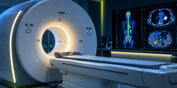
Having an MRI scan for cancer? Learn what it can show, how to prepare, what contrast involves and which questions to ask before your appointment.
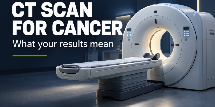
CT Scan for Cancer: What It Shows and What the Results Mean
CancerSnoop Test Guide
A clearer picture—not the whole story.
A CT scan for cancer can help locate abnormalities, assess disease extent and monitor treatment. Understanding the result means looking beyond a single measurement.
What does a CT scan show?
Computed tomography (CT) uses X-rays and computer processing to create cross-sectional images of the body. It shows anatomy: the location, size and appearance of organs and other structures.
In cancer care, CT can support diagnosis, staging, biopsy planning and follow-up. The body areas scanned and use of contrast depend on the clinical question. [1]
What Evidence Supports
An established imaging tool
CT can provide information that helps guide cancer care. Its usefulness depends on choosing the right examination for the question being asked.
Misleading Assumption
“One scan answers everything”
A report needs clinical interpretation. Ask what the findings establish, what remains uncertain and whether another investigation is needed.
Why is contrast sometimes used?
Contrast improves the visibility of selected tissues and blood vessels. CT may use injected iodinated contrast, oral contrast or neither, depending on the examination.
Before injected contrast, tell the imaging team about previous contrast reactions and kidney problems. Kidney-function testing may be needed. Follow the department’s preparation instructions rather than changing medicines yourself.
Allergic-like reactions to modern iodinated contrast are uncommon; severe reactions are rare. Kidney-related precautions depend on the patient’s condition, particularly acute kidney injury or severe kidney impairment. A kidney problem occurring after contrast is not automatically caused by it. [2]
Potential Harm
Report a reaction immediately
Tell staff immediately about breathing difficulty, throat swelling or other concerning symptoms during or after contrast administration. Severe symptoms after leaving the department require emergency medical attention. [2]
How should you read a CT report?
Use the report as a starting point for a discussion—not as a diagnosis to interpret alone. Useful questions include:
- Which findings matter for my care?
- Which are confirmed, and which are only suspected?
- What earlier scan was used for comparison?
- Does anything need follow-up, another imaging test or a biopsy?
- Does this result change the treatment plan?
If wording is unclear, ask your team to explain the exact sentence. Avoid drawing conclusions from an isolated phrase or measurement.
What do response, stable disease and progression mean?
Standardized response criteria help assess changes during treatment. RECIST 1.1 is a widely used framework for solid tumors, developed primarily for clinical trials. It evaluates selected lesions, other disease findings and new lesions—not just one measurement. [3]
- Partial response: sufficient reduction to meet the applicable criteria.
- Stable disease: neither sufficient shrinkage for response nor sufficient worsening for progression.
- Progressive disease: worsening that meets the relevant criteria.
Small measurement differences can reflect variability. Some treatments also change tumor density or internal structure without a matching change in size. Certain cancers and therapies require different assessment approaches. [3]
Interpretation Matters
Ask what “stable” means for you
Rather than treating the word as good or bad by itself, ask how it relates to your treatment goals, symptoms and previous scans.
Can a CT scan for cancer miss disease?
CT has detection limits. A reassuring scan is not a guarantee that microscopic disease is absent, and an abnormality does not necessarily establish cancer. The next step depends on what was seen and the clinical context. [1]
CT versus PET/CT
CT mainly provides structural information. PET uses a radioactive tracer to provide functional information; PET/CT combines the two. They answer related but different questions. [1]
Read our guide to PET scans in cancer.
What about radiation exposure?
CT uses ionizing radiation. A medically justified examination balances its expected benefit against radiation risk. Tell the imaging team if pregnancy is possible. Ask why each scan is needed rather than assuming either that every scan is harmless or that necessary imaging should be avoided. [1]
Before your next appointment
- Bring your questions and the exact wording you want explained.
- Ask whether the team has access to your previous images.
- Clarify who will discuss the result with you and when.
- Ask what happens next and which symptoms should prompt earlier contact.
CancerSnoop Bottom Line
Understand the finding. Ask about the next step.
A CT result is most useful when your team explains what it means for your care. You should leave that conversation knowing what is established, what remains uncertain and what happens next.
Related CancerSnoop Guides
- PET Scan in Cancer: What It Shows and What the Results Mean
- CEA Tumor Marker: What Does It Really Tell You?
- Explore Tests & Results
Scientific Sources
[1] National Cancer Institute — Computed Tomography (CT) Scans and Cancer
Institutional patient guidance. Reviewed February 8, 2024.
[2] American College of Radiology — ACR Manual on Contrast Media
2026 edition. Guidance on contrast administration, reactions and kidney-related precautions.
[3] Zamboni GA, et al. — ESR Essentials: response assessment criteria in oncologic imaging—practice recommendations by the European Society of Oncologic Imaging
European Radiology. 2025;35:674–683. Published online August 13, 2024.
DOI: 10.1007/s00330-024-11006-w
Educational information, not individual medical advice. Your medical team should interpret imaging findings in the context of your clinical situation.
Evidence checked: October 20, 2026. This date does not indicate an independent physician review.
Continue Exploring